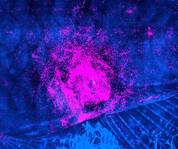
Immune cells called neutrophils (pink) aggregate to promote wound healing in a zebrafish model.
Credit: L. Greenspan, Weinstein Lab
A better understanding of the different phases of wound healing and how they are altered by aging or disease will help scientists develop new therapeutics to improve poor healing. Zebrafish offer researchers the opportunity to visualize and experimentally manipulate the cellular and molecular events that occur during wound healing. However, current methods to generate skin wounds in zebrafish require specialized equipment or yield inconsistent results.
- The Weinstein Lab developed an inexpensive, simple, and effective method for generating reproducible skin injuries in adult zebrafish. Their method uses a rotary tool to create small wounds on anesthetized zebrafish.
- By combining this new injury system with high-resolution live imaging, the scientists were able to monitor closing of the wound, immune cell recruitment and activation, and blood vessel regrowth in the same fish over time.
- The investigators published a detailed description of their experimental platform, allowing other scientists to adapt it to gain new insights into the cell biology of wound healing. The method can be applied to observe wound healing in real-time in transgenic zebrafish, allowing researchers to carefully study the cellular and molecular processes driving wound healing.
Reference
Greenspan LJ, Ameyaw KK, Castranova D, Mertus CA, Weinstein BM. Live imaging of cutaneous wound healing after rotary tool injury in zebrafish. J Invest Dermatol DOI: 10.1016/j.jid.2023.10.015 (2024)
Learn more about the Aquatic Models of Human Development Group: https://www.nichd.nih.gov/about/org/dir/affinity-groups/AMHD
 BACK TO TOP