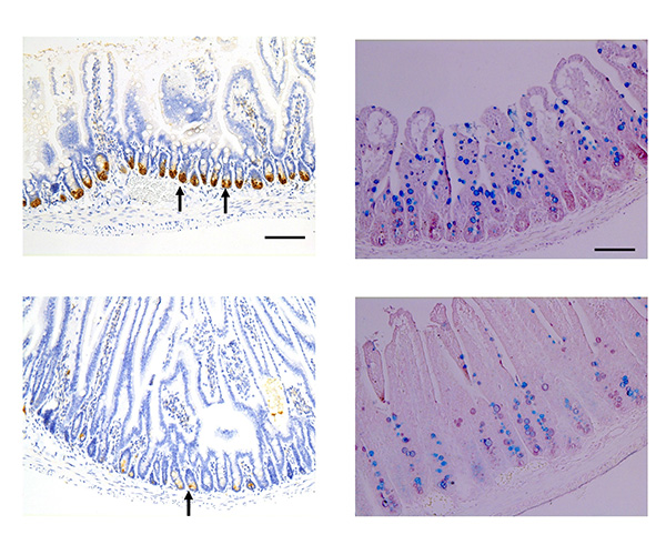
Top panels show sections of typical mouse intestine, and bottom panels show sections of intestine lacking SLC7A5. Those lacking SLC7A5 have fewer Paneth cells (left panels, brown, indicated with arrows) but more goblet cells (right panels, blue) in the crypts. In the absence of SLC7A5, goblet cells are reduced in the villi.
Credit: Shi Lab
The intestines digest and absorb nutrients and water, as well as form a protective barrier against harmful materials and microbes. Maintaining intestinal integrity and function in the face of a constant barrage of chemicals and microorganisms is critical for health. This requires a careful balance of interactions among different cell types within the intestinal lining (epithelium), the underlying tissues, the intestinal microbiota, and the immune system. Intestinal epithelial cells are continually renewed, developing from adult intestinal stem cells into specialized cell types—a process known as differentiation.
- Many genes and cell-signaling networks can influence the proliferation of intestinal epithelial cells. Among these is mTORC1, activation of which increases cell proliferation. Using mouse models, the Shi Lab deleted SLC7A5—a transporter protein capable of activating mTORC1—from intestinal epithelial cells.
- Loss of SLC7A5 in the intestinal epithelium reduced mTORC1 signaling and dramatically decreased the overall number of mature secretory cells, including Paneth cells, which secrete anti-microbial proteins. Numbers of goblet cells, which secrete mucus that coats the epithelium, were lower in the villi—finger-like projections that line the intestine—but higher in the areas between villi, called crypts.
- The researchers found increased cell proliferation in the crypts, but lower numbers of mature Paneth cells. Further experiments revealed that Paneth cells underwent dedifferentiation (a process in which they became less specialized), leading to fewer mature cells with little effect on overall Paneth cell number.
- The findings suggest that SLC7A5 regulates secretory cell differentiation and indirectly regulates cell proliferation, highlighting the complex interactions needed to maintain the intestine’s physiological status.
Reference
Bao L, Fu L, Su Y, Chen Z, Peng Z, Sun L, Gonzalez FJ, Wu C, Zhang H, Shi B, Shi YB. Amino acid transporter SLC7A5 regulates cell proliferation and secretory cell differentiation and distribution in the mouse intestine. Int J Biol Sci DOI: 10.7150/ijbs.94297 (2024)
Learn more about the Cell Regulation and Development group: https://www.nichd.nih.gov/about/org/dir/affinity-groups/CRD
 BACK TO TOP