Exploring Links Between Genetic Ancestry and Fetal Growth
NICHD researchers study various factors that influence healthy pregnancies and optimal outcomes. Learn about findings from the Epidemiology Branch on how genetic ancestry influences fetal growth and maternal risk for type 2 diabetes.
Unraveling Causes of Primary Ovarian Insufficiency
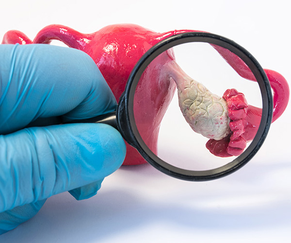
Many girls and women with galactosemia, an inherited condition in which the body is unable to metabolize the simple sugar galactose, develop primary ovarian insufficiency. Learn about work from the Pediatric and Adolescent Gynecology Program to understand the underlying mechanisms.
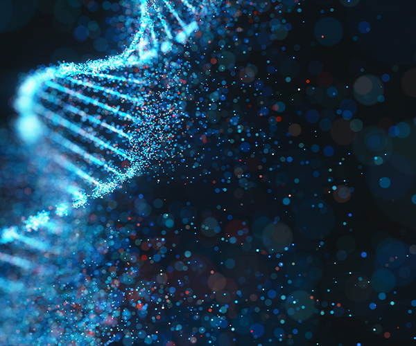
Discovering How Chromosomes Pair During Sperm Formation
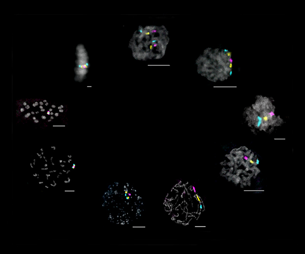
During the creation of egg or sperm cells—a specialized cell division called meiosis—maternal and paternal chromosome copies need to find each other for genetic recombination before the cell divides. Learn about work from the Rosin Lab on how this process is regulated in sperm.
Evaluating Gene Editing to Treat a Rare Disease
People with glycogen storage disease type Ia (GSD-Ia) cannot maintain blood sugar levels, causing seizures, growth problems, and other issues. Learn about work from the Chou Lab to correct the genetic variation that underlies GSD-Ia.
Understanding How Microglia Develop in the Brain
Microglia are a type of immune cell found in the brain, and these cells are responsible for development and maintenance of a healthy brain throughout life. Learn about work from the Ozato Lab on understanding the genetic programs underlying microglia development.
Probing the Cell’s Protein-Sorting Network
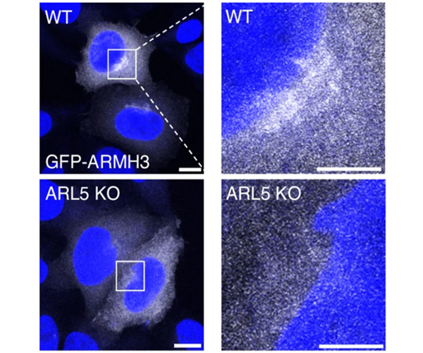
ARL5 works as a “molecular switch” at the cell’s protein-distribution center by regulating protein modifications and retrieval of recycled transport machinery. Learn about work by the Bonifacino Lab that expands upon ARL5’s function and interacting partners.
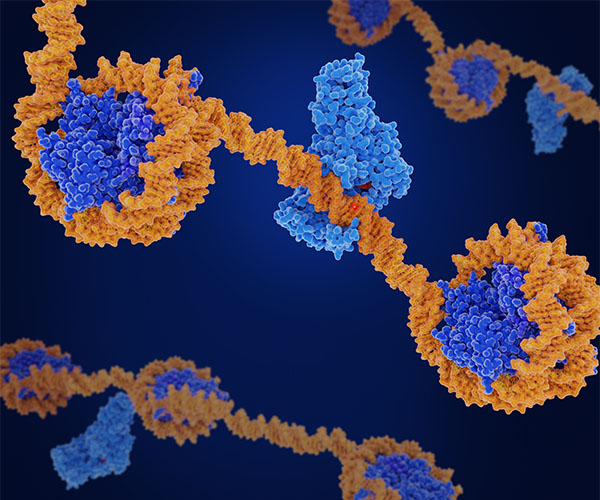
Discovering Critical Roles for Sequences that Set the 3D Structure of DNA
Genes can be regulated at the physical level, where structural loops are able to recruit or impair DNA transcription. Learn about work from the Rocha Lab on how disruption of small regulatory sequences responsible for these loops can have outsized effects on development.
Illuminating Early Indicators of Suicide Vulnerability

Growing evidence suggests that vulnerability to suicide is partly established early in life. Learn about work from the Social and Behavioral Sciences Branch assessing these early indicators.
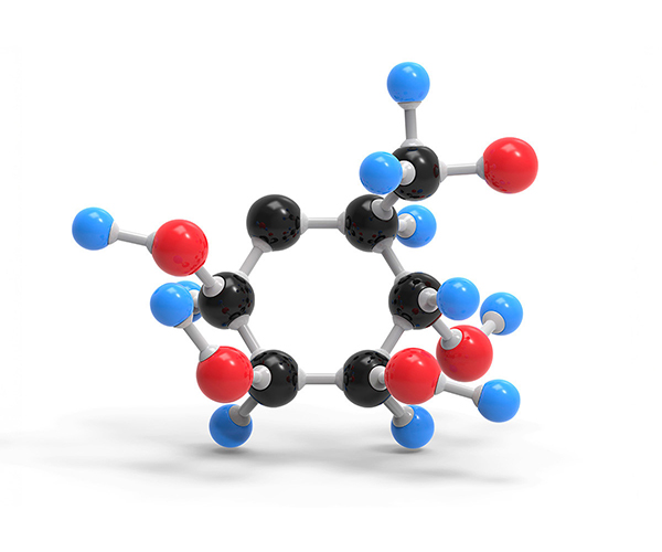
Linking the Role of Iron-Sulfur Enzymes to Human Health and Disease
Iron is an essential trace element for all living organisms and plays a crucial role in various physiological processes. This activity relies on iron-sulfur enzymes and related proteins within cells. Learn about work from the Rouault Lab on a disorder caused by mutations in the CIAO1 gene that affects iron-sulfur proteins.
Developing New Methods to Image and Reconstitute Complex Proteins
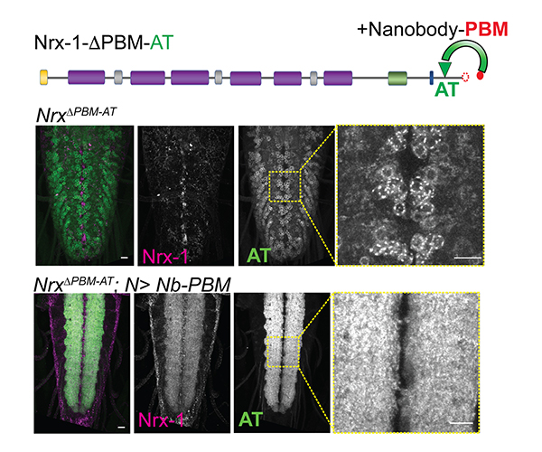
Neurexins are complex proteins critical for the formation of synapses, which connect neurons and help transmit signals. Learn about work from the Serpe Lab on a new method to image, track, and reconstitute neurexins.
Understanding How Lyme Bacteria Move
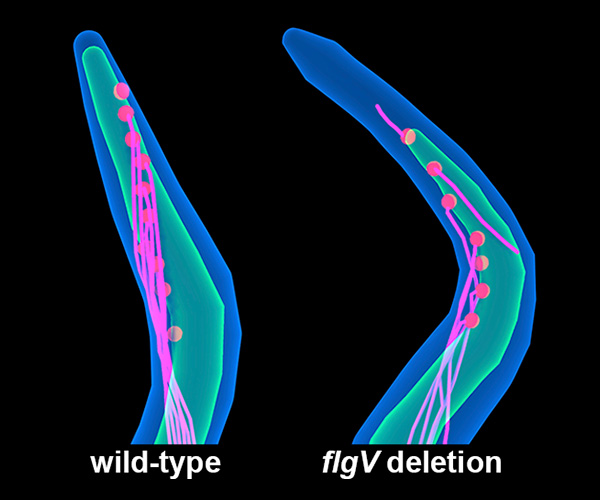
Understanding how Lyme bacteria move, replicate, and divide inside their hosts provides key insights into the biology of the disease. Learn about work from NICHD researchers to understand the regulation of whiplike structures that help bacteria move.
Back to Research Highlights from the Division of Intramural Research.
 BACK TO TOP