
Image collection promises to inform patient care and studies of placental disorders
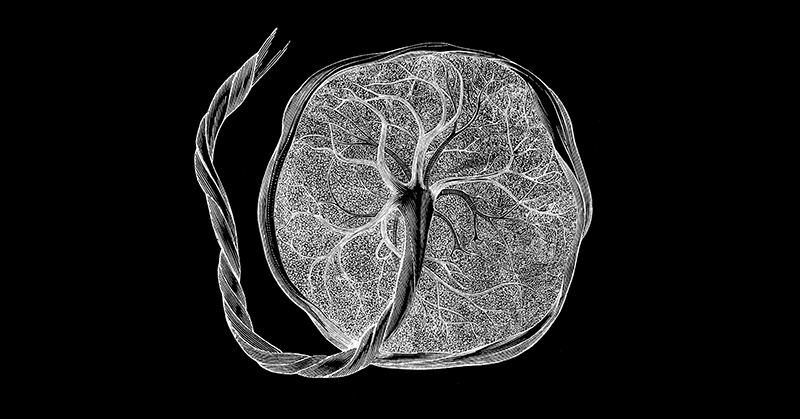
Using new ultrasound technology that can visualize and measure blood flow through extremely small blood vessels, researchers funded by the National Institutes of Health have tracked the placenta’s development from the merging of placental cells with blood vessels in the uterus to a fully formed organ. The images and data are taken from the 12th through 37th week of pregnancy and provide a reference for normal placental development to help with comparison studies, tracking of placental development in healthy pregnancies, and identification of placental disorders that may lead to low birth weight, preterm birth, and other complications.
The study was conducted by Rebecca Horgan, M.D., of the Eastern Virginia Medical School, and colleagues. It appears in Ultrasound in Obstetrics & Gynecology. Funding was provided by NIH’s Eunice Kennedy Shriver National Institute of Child Health and Human Development.
Background
Early in pregnancy, cells from the placenta invade the spiral arteries in the uterus, making them wider and reducing resistance in the vessel walls. This remodeling process increases the blood supply available to the placenta, allowing it and the fetus to continue growing until birth. Failure of spiral arteries to widen sufficiently is thought to result in an underdeveloped placenta, depriving the fetus of oxygen and nutrients, interfering with its growth, and resulting in complications such as preterm birth or infants being born small for their gestational age. This failure also may lead to preeclampsia, a potentially life-threatening complication of pregnancy resulting in high blood pressure and protein in the urine.
Previous ultrasound techniques can depict larger uterine arteries, but they aren’t sensitive enough to measure blood flow in small blood vessels like the spiral arteries. For the current study, researchers used Superb Micro-Vascular Imaging® (SMI), a recently developed ultrasound technology capable of imaging microscopic blood vessels.
Results
Researchers enrolled 90 pregnant people in the study. Beginning in the 12th to 13th week of pregnancy, ultrasound images of the placenta were taken every two weeks until the 16th week of pregnancy and then every 4 weeks until 37 weeks of pregnancy, resulting in 8 ultrasound exams per pregnancy. Researchers took precise measurements of the spiral arteries at the location where they opened into the intervillous space—the channel between maternal and fetal parts of the placenta, where fetal cells access maternal blood.
Significance
Although previous studies have used ultrasound to visualize and measure flow through spiral arteries, the authors said theirs is the first study to do so in the first trimester. The images and measurements will provide standards of normal placental development to inform studies of preeclampsia and other conditions affecting placental development.
Reference
Horgan, R, et al. Longitudinal assessment of spiral and uterine arteries in normal pregnancies using novel ultrasound tools. Ultrasound in Obstetrics & Gynecology. 2023.
 BACK TO TOP